Skip to main content Skip to navigation

Linear Dichroism

Why do we use linear dichroism?

Linear Dichroism (LD) is a spectroscopic technique that can be used with systems that are either intrinsically oriented, or can be oriented during an experiment by external forces. It gives information about conformation and orientation of structures within molecules.

Knowing the structure of a molecule is one of the keys to deducing its function in a biological system. Many biomacromolecules, however, are not amenable to structural characterisation by powerful techniques such as NMR and X-ray diffraction because they are too large, too flexible or cannot be crystallized. Long molecules such as

DNA, DNA-ligand complexes, DNA-enzyme complexes

Fibrous proteins including cytoskeletal proteins, Alzheimers and prion proteins, collagen

Membrane proteins and peptides in liposomes (which are distorted by the flow so acquiring a high enough aspect ratio to orient)

Carbon nanotube, carbon nanotube-ligand complexes

LD is also ideal for following the kinetics of any reaction which involves changing the structure or length of a molecule. A simple example is that of restriction enzyme activity on DNA.

What is linear dichroism?

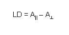
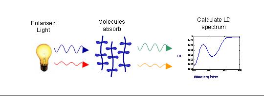
To measure LD the sample is oriented, then the difference in absorption of light linearly polarised parallel and perpendicular to the orientation axis is measured

ld_eqn.jpg

In the case of flow orientation of long molecules the orientation axis is the long axis of the molecule. The figure below shows a schematic of how LD is measured.

LD schematic

We have designed the Couette flow LD cells we use in our research. The refinements of our original designs are now made by Diotica Scientific Ltd. and sold by Kromatek http://www.kromatek.co.uk/categories/Linear-Dichroism-Couettes

Representative publications

Rodger, A.; Nordén, B. “Circular dichroism and linear dichroism”; Oxford University Press, 1997, pp150 (unfortunately this is out of print, but we are working on a new edition)

Bulheller, B.; Rodger, A.; Hirst, J.D. “Circular and linear dichroism of proteins” Physical Chemistry Chemical Physics, 2007, 9, 2020-2035

Small, E.; Marrington, R.; Rodger, A.; Scott, D.J.; Sloan. K.; Roper, D.; Dafforn, T.R.; Addinall, S.G. “FtsZ polymer-bundling by the Escherichia coli ZapA orthologue, YgfE involves a conformational change in bound GTP” Journal of Molecular Biology, 2007, 369, 211-221

Rodger, A.; Marrington, R.; Geeves, M.A.; Hicks, M.; de Alwis, L.; Halsall, D.J.; Dafforn, T.R. “Looking at long molecules in solution: what happens when they are subjected to Couette flow?” Physical Chemistry Chemical Physics, 2006, 8, 3161-3171

Hicks, M.R.; Rodger, A.; Thomas, C.M.; Batt, S.M.; Dafforn, T.R. “Restriction enzyme kinetics monitored by UV linear dichroism” Biochemistry 2006, 45, 8912-8917

Marrington, R.; Seymour, M.; Rodger, A. “A new method for fibrous protein analysis illustrated by application to tubulin microtubule polymerisation and depolymerisation” Chirality 2006, 18, 680-690

Rajendra, J.; Damianoglou, A.; Hicks, M.; Booth, P.; Rodger, P.M.; Rodger, A. “Quantitation of protein orientation in flow-oriented unilamellar liposomes by linear dichroism”, Chemical Physics 2006, 326, 210–220

Marrington, R.; Dafforn, T.R.; Halsall, D.J.; Hicks, M.; Rodger, A. “Validation of new microvolume Couette flow linear dichroism cells” Analyst, 2005, 130, 1608-1616

Rajendra, J.; Rodger, A. “The binding of single stranded DNA and PNA to single walled carbon nanotubes probed by flow linear dichroism” Chemistry - A European Journal 2005, 11, 4841-48

Halsall, D.J.; Dafforn, T.R; Marrington, R.; Halligan, E.; and Rodger, A. “Linear Dichroism for quantitative Polymerase Chain Reaction applications: An intrinsic signal from DNA has the potential to be an interference-free, specific and label free probe for real-time and quantitative PCR” IVD Technology, 2004, 6, 51-60

Dafforn, T.R. and Rodger, A. “Unravelling the configuration of protein fibres and membrane proteins” Current Opinion in Structural Biology, 2004, 14, 541-546

Marrington, R.; Small, E.; Rodger, A.; Dafforn, T.R.; Addinall, S. “FtsZ fibre bundling is triggered by a calcium-induced conformational change in bound GTP” Journal of Biological Chemistry 2004, 47, 48821-48829

Marrington, R.; Dafforn, T. R.; Halsall, D.J.; Rodger, A. “Micro volume Couette flow sample orientation for absorbance and fluorescence linear dichroism” Biophysical Journal, 2004, 87, 2002-2012

Rajendra, J.; Baxendale, M.; Dit Rap, L.G.; Rodger, A. “Flow linear dichroism to probe binding of aromatic molecules and DNA to single walled carbon nanotubes” Journal of the American Chemical Society, 2004, 126, 11182-11188

LD cell


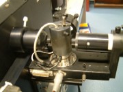
 A Couette flow cell used for LD.

The light beam comes from the left and is collected by a photomultiplier tube on the right, having passed through the sample.

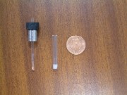
Below is a sample holder and rod.


Cell and capillary, small version