Biological Systems
|
|
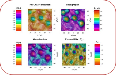
Scanning electrochemical microscopy is widely used to investigate various aspects of biological systems such as morphology, metabolism and transport of different signalling molecules. Our research mainly focusses on electrochemical probing of cell surfaces, development of integrated electrochemical microscopy techniques capable of monitoring and investigating specific metabolites and biological reactions with high spatial resolution and investigation of material transport across biological membranes. A broad range of electrochemical scanned probe techniques are used including SECM, SICM, combined SECM-SICM, IC-SECM and SECCM, These scanning electrochemical microscopy techniques were also integrated with optical microscopy systems such as ultra-fast confocal laser scanning microscopy and other related technologies, many of which are pioneered in our group. |
|
|
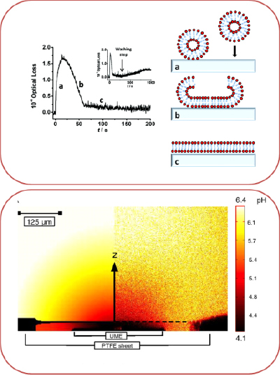
Supported lipid bilayer systems were extensively used as model cellular membranes for the fundamental studies of biophysical processes. A key aspect of our research in this area is to probe and confirm formation of supported lipid bilayer and subsequently study dynamic processes taking place on and across this membrane. Transport of molecules across cell membrane is fundamental processes in cellular biology. They are also becoming increasingly important in many medical, pharmaceutical, and environmental technologies. Our research mainly focusses on development of new method to investigate the formation of supported bilayers on various solid substrate systems by using techniques like AFM, evanescent wave cavity ring down spectroscopy and quantitatively measure the permeation of various molecules via microelectrochemistry, combined with laser confocal scanning microscopy (LCSM) and supported by finite element modeling (FEM).
|
|
|
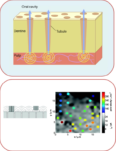
Dentinal hypersensitivity is a common problem, affecting much of the adult population. It is caused by the movement of fluid through exposed dentinal tubules. Scanning Electrochemical Microscopy (SECM) has been shown to be a powerful technique for the measurement of local solution velocities through human dentine slices, in-vitro, and for assessing quantitatively the effect of treatments on fluid flow. The main goal of our research is to improve the spatial resolution and contrast of SECM measurements of transport through model porous materials by using improved electrochemical techniques like intermittent contact (IC)-SECM, which will open a new way to understand and treat dentinal hypersensitivity.
|
|
|
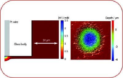
The rate of dental enamel dissolution is very important in the area of dental science in the context of acid erosion and in understanding how this process can be inhibited. In our group, we use scanning electrochemical microscopy (SECM) for the quantitative study of acid-induced bovine enamel dissolution. High rates of mass transport in SECM allow the surface kinetics of dissolution to be evaluated. SECM is able to deliver high, controllable, and local acid challenges in a defined way and that multiple dissolution measurements can be performed on one sample, eliminating intersample variability effects.
|