Skip to main content
Skip to navigation
Sign in
Study
Research
Business
Alumni
News
Engagement
Search Warwick
Search
Warwick Systems Biology Centre
About Us
Find Us
Contact Us
Research
Research Areas
Publications
Computing Facilities
Software Downloads
Past research
Staff
Administration
Piotr Baniukiewicz
Alina Bazarova
Till Bretschneider
Nigel Burroughs
Giorgos Minas
Hiroshi Momiji
Jay Moore
David Rand
Graham Wood
Students
Events
SBIDER calendar
News
Intranet
(Restricted permissions)
Info & Forms
(Restricted permissions)
Internal talks
(Restricted permissions)
SBIDER Seminars
(Restricted permissions)
WSB Archive
(Restricted permissions)
Staff
Till Bretschneider
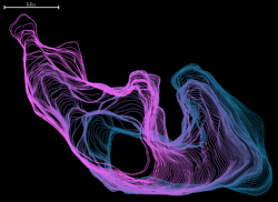
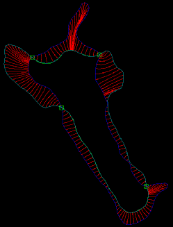
QuimP Software
QuimP Software
QuimP has moved to
http://warwick.ac.uk/quimp